


Medical Articles
by William H. Bates, M. D. The Bates Method is a method to restore eyesight naturally-without the use of glasses, contact lenses, surgery or drugs.
Reprinted from New York Medical Journal, March 16, 1912, pp. 529-532. Read before the New York Country, Medical Association, January 23, 1912
THE CAUSE OF MYOPIA.
By W. H. BATES, M. D.,
New York
In the normal eye parallel rays of light are focused on the retina; in myopia they are focused in front of the retina. Myopia, with elongation of the optic axis from bulging of the posterior pole, posterior staphyloma, is incurable. Rarely congenital, myopia is usually acquired.
Functional myopia is an early stage of myopia with elongation of the eyeball. It is produced by muscular action, which alters the curvature of the crystalline lens, modifies the convexity of the cornea, or produces an elongation of the eyeball. Voluntary functional myopia may be produced by efforts to see distant objects, in children, elderly people, cases in which the accommodation is apparently paralyzed by atropine, and in aphakia after cataract extraction. That muscular action can produce functional myopia is shown by the fact that many cases of voluntary functional myopia manifest a convergent, divergent, or vertical squint. Also, operations on the eye muscles have benefited functional myopia. Von Graefe, Donders, and others have reported good results in functional myopia after tenotomy of the external rectus. Stevens published (Anomalies of the Eye Muscles) some cases of functional myopia relieved after operations on the eye muscles. In a personal communication he said that in his experience the refraction of the eye was usually changed after such operations.
The diagnosis of myopia may be made with the ophthalmoscope or retinoscope. In myopia with elongation of the eyeball, with the ophthalmoscope by the direct method, the details of the fundus cannot be seen clearly without the aid of a concave glass; whereas, in functional myopia, the retinal vessels and chorioidal pigment can be seen clearly, occasionally without such a glass. With the retinoscope, in myopia with elongation of the eyeball the shadow seen with the plane mirror held at four feet or further always moves in the opposite direction to the movements of the mirror; but, in functional myopia the shadow moves in the same direction at times, and especially when the eye is regarding distant objects without especially trying to see.
It has been generally accepted, that after the prolonged use of atropine, if the myopia continues, it is due to permanent elongation of the eyeball. After twenty-five, years' study of these cases, my experience leads me to the conclusion that atropine does not always relax the near focus or relieve functional myopia.
A study of the eyes of a large number of individuals in whom functional myopia was produced by an effort, unconsciously or voluntarily, may be briefly summarized as follows:
An unsuccessful effort of the normal eye to see accurately new, strange, or unfamiliar distant objects was always followed either by myopic astigmatism, usually—compound myopic astigmatism, occasionally, or simple myopia infrequently. Mixed astigmatism was not observed. For example:
Case I. A woman, aged twenty-five years, had diffi-culty in reading the ten line of the Snellen card at ten feet. When she was unable to see the letters, retinoscopy always indicated a myopic refraction; but, when she read the letters, simultaneous retinoscopy always indicated no myopia. So accurate was retinoscopy in measuring the refraction that one was invariably correct when telling her when she could see and when not.
Case II. A boy, aged nine years, while reading at ten feet the line marked ten on the Snellen card was not myopic. When he regarded the large letter, vision 10/200, he had myopic astigmatism When he regarded a picture at twenty feet, he appeared to make a greater effort to see, and by simultaneous retinoscopy, he had compound myopic astigmatism.
Case III. A boy, aged five years, when regarding his mother at ten feet, by retinoscopy was not myopic; but, when he regarded a stranger at ten feet, or the unknown letters on the Snellen card at the same distance, he had myopic astigmatism. When he made a manifestly increased effort to see a dog at 100 feet, the objective test used simultaneously indicated compound myopic astigmatism. The increased effort to see distant objects produced more myopic refraction.
Case IV. A woman, aged thirty-six years, with vision, 10/200, 10/50, 10/10, was not myopic. Neither was she myopic when she regarded at ten feet or 100 feet a picture, a book, and many other objects; but, when she was asked to look directly at a point three feet to one side of the Snellen card and read the letters, which was impossible, the retinoscope indicated compound myopic astigmatism, and tie left eve converged. (Figs. 1 and 2.)
Case V. A girl aged eighteen years, emmetropic, was similar to the previous patient; she did not make an effort to see distant objects until asked to regard the Snellen card by excentric fixation. Compound myopic astigmatism was produced and the right eye diverged.
Case VI. A man, aged twenty years, had used atropine sulphate, one per cent., three times a day, In the left eye for two months. When he regarded a green curtain at ten feet he was not myopic; but, when regarding the large letters on the Snellen card he had compound myopic astigmatism.
Cass VII. A woman, aged forty-seven years, right eye, keratoiritis, received atropine sulphate, one per cent., three times a day for fifty days. When she regarded a green curtain at ten feet, she was not myopic; but, when she read some of the large letters on the Snellen card at ten feet, retinoscopy indicated compound myopic astigmatism.
Case VIII. A man aged seventy years, by retinoscopy was not myopic when reading the ten line at ten feet; but, when he regarded an indistinct object, a thermometer, at 100 feet, retinoscopy indicated myopic astigmatism. An increased effort produced compound myopic astigmatism.
In normal eyes the axis of myopic astigmatism, which was found by retinoscopy after an effort to see distant objects, was usually corrected by a concave cylinder at 180°. It was observed frequently at 90°, and less often in an oblique meridian. As a rule the vertical or horizontal axis was the same in each eye—exceptions were found infrequently. When the axis was oblique in one eye it was generally parallel, or else at right angles, in the other eye. In most individuals the axis was always the same when tested frequently, daily, weekly, or after some months. Occasionally the axis would change in one person from 90° to 180°, or the reverse, or became more or less oblique when making apparently the same effort to see distant objects. The maximum amount of myopic astigmatism produced was 4 D., and was observed in a man aged fifty-nine years, with normal eyes when he regarded an astigmatic chart at ten feet.
In most eyes with errors of refraction, and in normal eyes with excentric fixation, the axes of astigmatism produced by efforts to see distant objects were not usually constant, and greater variations occurred in the same eye from day to day than was observed in normal eyes. In compound hypermetropic astigmatism the effort to see at a distance always lessened the refraction of sometimes one, sometimes the other principal meridian, or of both. In compound myopic astigmatism, one or both of the principal meridians were always increased. In mixed astigmatism, sometimes the hypermetropic meridian was lessened; in other cases the myopic meridian was increased, and in still others the hypermetropic meridian was lessened, while the myopic meridian was increased.
Symptoms of effort when trying to see distant objects: School children and others usually showed by facial expression that an effort was made—the eyelids were partly closed, or the reverse, more open, staring; wrinkling of the skin of the forehead and eyelids, contortions of the facial muscles, inclinations of the head in various directions; tremor of the head, and movements of the eyeballs resembling nystagmus were observed. Many school children and adults with normal eyes produced temporary excentrtc fixation, either with convergent, divergent, or vertical squint when trying unsuccessfully to read the Snellen card. The eyes of more than 10,000 school children were examined during the past ten years. The efforts of many to see were so manifest that one could usually tell before the sight was tested that their vision was defective (Figs. 3, 4, 5).
Recently a public school in New York was visited. In one class room of thirty young pupils, the attention of the principal was directed to five children whose facial expression suggested defective vision. She tested their sight and found it poor in all. She proposed glasses. In a few minutes the children were shown how to read the small letters on the Snellen card. They obtained normal vision and required no glasses. The facial wrinkles and evidences of strain disappeared.
About twenty-five teachers listened to a talk on myopia. Most of them showed by their facial expression, wrinkles of the forehead, and strained look of their eyes that their vision was probably defective. They were recommended to read the small letters on the Snellen card. The majority obtained normal vision almost immediately; the wrinkles were lost, and their eyes and faces no longer had the appearance of strain.
H. Cohn (The Hygiene of the Eye in Schools, p. 53) wrote: "All oculists agree that protracted near work with a bad light is one of the circumstances most favorable to the origin and development of short sight." My observations did not support this statement.
The near focus of the normal eye was measured objectively with the aid of the retinoscope. When a normal eye read fine print, diamond type, Jaegar No. 1, readily, without effort, at twelve inches, a concave twelve inch glass held outside the visual axis corrected the focus. When the eye read at ten inches it was too weak to correct the focus; and when the print was read at a greater distance than twelve inches, the glass was too strong, over-corrected the focus. Retinoscopy always measured the focus accurately and simultaneously while the normal eye read at 6", 10", 20", 40", or at any distance the fine print.
When the illumination of the print was lessened sufficiently to make it difficult to read Jaeger 1 at twelve inches, retinoscopy indicated that the near focus of the eye was not increased, but lessened in one or all meridians. No exceptions were found. It occurred in all school children, adults, and elderly people with normal eyes. Usually only one meridian was lessened, the horizontal. The maximum amount was 3 D. The vertical meridian was lessened, exceptionally.
Patients with emmetropia or normal refraction under atropine were examined. When large print was read, easily at twelve inches, the eye was focused as in distant vision; but when, because of less light, or the request to read smaller print, an effort was made, one or all of the principal meridians became hypermetropic. It was interesting to note that these same individuals always produced myopic refraction, usually greater in the horizontal meridian, while malting an effort to see distant objects; when an effort to see near always produced the opposite refraction, hypermetropia, and greater in the same meridian, the horizontal.
In hypermetropia, with or without astigmatism, one or more of the meridians of the eye were increased by efforts to read by a dim light. In myopia, with or without astigmatism, one or more of the meridians became less, myopic. In mixed astigmatism the refraction of the horizontal meridian became either less myopic or more hypermetropic when an effort was made to read fine print In presbyopia no exceptions were found; an effort to read always produced hypermetropia in one meridian in normal eyes, increased it in hypermetropia, or diminished it in myopia. In diseased conditions, inflammations of the eyelids, cornea, iris, retina, chorioid, and in cataract, an effort to read always lessened the focus.
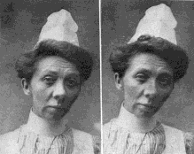
Fig. 1.—Reading the Snellen test card with normal vision; optic axesparallel.
Fig. 2.—The same patient making an effort to see the Snellen test card at ten feet by excentric fixation. The patient produced a functional myopia and the left eye turned in.
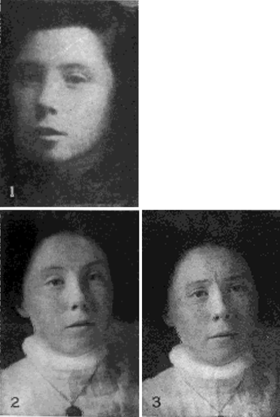
Fig. 3.—Girl with normal vision in 1904. Note the absence of facial effort.
Fig. 4.—The same girl as shown in Fig. 3, five years later with myopia of 3.00 D. Note the elevation of the eyebrows and other manifestations of effort.
Fig. 5.—The same, with myopia increased by voluntary effort to see better the Snellen card at twenty feet. The manifestation of effort is increased.
So decided was the relaxation of the near focus that efforts to read by a dim light were successfully employed in some cases of functional myopia to obtain adjustment of the eye for distant vision after other methods had failed.
The following cases illustrate the effects of effort when reading with difficulty at a near point:
Case IX. A boy with normal eyes, aged nine years, read jaeger No. 1 easily at twenty inches. A concave twenty inch glass held outside the visual axis corrected the focus in all meridians. When the light was lessened, the print was read with difficulty. Now retinoscopy indicated that the vertical meridian was accommodated as before, but the horizontal was lessened and had become hypermetropic. With the aid of the retinoscope one always knew when the boy read easily or with difficulty, He was also examined after he had been reading two hours by a poor light, leaning over, the book held in his lap. The result was the same.
Cask X. A girl, aged twelve years, compound hypermetropic astigmatism, left vision, 10/10 nearly. Retinoscopy, vertical meridian was corrected by convex 3.00 D. and the horizontal by convex 1.50 D. when she regarded the 200 Snellen card letter at ten feet. When she read the twenty line Snellen at ten inches the vertical meridian was corrected by a concave ten inch glass and the horizontal by concave 1.00 D. She read Jaeger No. 1 at ten inches with difficulty; the vertical meridian remained the same, while the horizontal was corrected by convex 1.00 D. The illumination of the page was reduced by a screen. She had greater difficulty in reading Jaeger No. 1 at ten inches when the retinoscope used simultaneously indicated that the vertical meridian was corrected by concave 4. D., or ten inch, while the horizontal was corrected by convex 2.30 D. Retinoscopy indicated that this patient read with difficulty even very large print. An increased effort did not increase the myopic refraction of the vertical meridian, but made the horizontal more hypermetropic than when regarding the Snellen card at ten feet.
Case XI. A girl, aged seven years, left eye under atropine sulphate, one per cent., three times a day for two months, vision normal with convex 3,00 D. S. combined with convex 0.50 D. C, at 90°, the same refraction with retinoscopy. With her correction she read with difficulty large print, Jaeger No. 14, at six inches when the vertical meridian was corrected by convex 4.00 D. and the horizontal by convex 5.00 D., an increase of 2. D. of hypermetropia in the horizontal meridian after an effort to read with the accommodation apparently paralyzed by atropine.
Case XII. A woman, aged seventy-six years, right eye, 20/30. no glass improved, incipient cataract. By retinoscopy all meridians were corrected by convex 0.50 D. Regarding the tip of her finger at six inches, the vertical meridian by retinoscopy was measured by convex 2.00 D., and the horizontal by convex 4.00 D. An effort to see distant objects always produced myopic refraction, while an effort to see near objects always produced the opposite, hypermetropic refraction.
Sufficient evidence has been obtained to convince me that near use of the eyes is not the cause of myopia. The cause of myopia is the same in birds, the lower animals, uncivilized man, and school children.
Wild birds have unusually good distant vision; but in captivity they acquire myopia (Casey A. Wood, Ophthalmology, Chicago, April, 1907). Uncivilized people have good sight; but after they live in civilized communities they acquire myopia (Risley, System of Diseases of the Eye, Norris & Oliver, 1897, Vol. II). Children in the first year of school have normal vision; later, myopia is acquired. The following explanation of these facts is offered:
The uncivilized man is compelled to adjust his eyes for accurate distant vision, for protection against enemies, and in obtaining food. But, when living in civilized communities he is protected from enemies, his food is supplied, accurate distant vision is no longer necessary, he neglects to practise it, naturally loses it, and becomes myopic. Wild birds are compelled to adjust their eyes accurately for distant vision; but, in captivity the necessity ceases, and, because accurate distant vision is no longer required, they neglect it and become myopic. School children do not need accurate adjustment of their eyes for distant vision. When they neglect to practise it they become myopic. To make the matter clearer: When the eyes are not accurately adjusted for distant vision they must obviously be adjusted for a near point and be functionally myopic
CONCLUSIONS.
1. Myopia is not caused by efforts to read by a bad light.
2. The cause of myopia is an effort, usually unconscious, to see distant objects.
938 St Nicholas Avenue.
| Eyes carePhysicianBate's booksLaser corre.Blues under eyesburning in the eyesanother diseasesMedical mistery Naturally eyesight correction. No laser eye surgery. Restore eyesight. Vision correction. Телефоны индивидуалок Питера. | |