


Perfect Sight Without Glasses
by William H. Bates, M.D.
Bates Method is a method to restore eyesight naturally-without useing of glasses, contact lenses, surgery or drugs.
PERFECT SIGHT WITHOUT GLASSES
CHAPTER IV
THE TRUTH ABOUT ACCOMMODATION AS DEMONSTRATED BY EXPERIMENTS ON THE EYE MUSCLES OF FISH, CATS, DOGS, RABBITS AND OTHER ANIMALS
THE function of the muscles on the outside of the eyeball, apart from that of turning the globe in its socket, has been a matter of much dispute; but after the supposed demonstration by Helmholtz that accommodation depends upon a change in the curvature of the lens, the possibility of their being concerned in the adjustment of the eye for vision at different distances, or in the production of errors of refraction, was dismissed as no longer worthy of serious consideration. "Before physiologists were acquainted with the changes in the dioptic system,"1 says Donders, "they often attached importance to the external muscles in the production of accommodation. Now that we know that accommodation depends on a change of form in the lens this opinion seems scarcely to need refutation." He states positively that "many instances occur where the accommodation is wholly destroyed by paralysis, without the external muscles being the least impeded in their action," and also that "some cases are on record of paralysis of all or nearly all of the muscles of the eye, and of deficiency of the same, without diminution of the power of accommodation."2
If Donders had not considered the question settled, he might have inquired more carefully into these cases, and if he had, he might have been less dogmatic in his statements; for, as has been pointed out in the preceding chapter, there are plenty of indications that the contrary is the case. In my own experiments upon the extrinsic eye muscles of fish, rabbits, cats, dogs and other animals, the demonstration seemed to be complete that in the eyes of these animals accommodation depends wholly upon the action of the extrinsic muscles and not at all upon the agency of the lens. By the manipulation of these muscles I was able to produce or prevent accommodation at will, to produce myopia, hypermetropia and astigmatism, or to prevent these conditions. Full details of these experiments will be found in the "Bulletin of the New York Zoological Society" for November, 1914 [link], and in the "New York Medical Journal" for May 8, 1915 [link]; and May 18, 1918 [link]; but for the benefit of those who have not the time or inclination to read these papers, their contents are summarized below.
There are six muscles on the outside of the eyeball, four known as the "recti" and two as the "obliques." The obliques form an almost complete belt around the middle of the eyeball, and are known, according to their position, as "superior" and "inferior." The recti are attached to the sclerotic, or outer coat of the eyeball, near the front, and pass directly over the top, bottom and sides of the globe to the back of the orbit, where they are attached to the bone round the edges of the hole through which the optic nerve passes. According to their position, they are known as the "superior," "inferior," "internal" and "external" recti. The obliques are the muscles of accommodation; the recti are concerned in the production of hypermetropia and astigmatism.
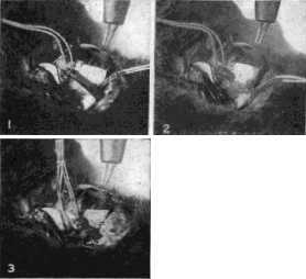
Fig. 13. Demonstration Upon the Eye of a Rabbit that the Inferior Oblique Muscle is an Essential Factor in Accommodation
No. 1.—The inferior oblique muscle has been exposed and two sutures are attached to it. Electrical stimulation of the eyeball produces accommodation as demonstrated by simultaneous retinoscopy.
No. 2.—The muscle has been cut. Electrical stimulation produces no accommodation.
No. 3.—The muscle has been sewed together. Electrical stimulation produces normal accommodation.
In some cases one of the obliques is absent or rudimentary, but when two of these muscles were present and active, accommodation, as measured by the objective test of retinoscopy, was always produced by electrical stimulation either of the eyeball, or of the nerves of accommodation near their origin in the brain. It was also produced by any manipulation of the obliques whereby their pull was increased. This was done by a tucking operation of one or both muscles, or by an advancement of the point at which they are attached to the sclerotic. When; one or more of the recti had been cut, the effect of operations increasing the pull of the obliques was intensified.
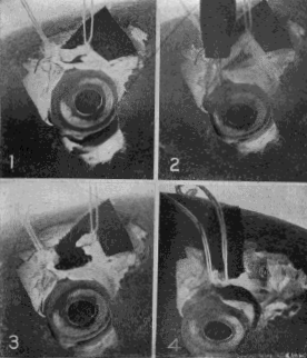
Fig. 14. Demonstration Upon the Eye of a Carp That the Superior Oblique Muscle Is Essential to Accommodation
No. 1.—The superior oblique is lifted from the eyeball by two sutures, and the retinoscope shows no error of refraction.
No. 2. — Electrical stimulation produces accommodation, as determined by the retinoscope.
No. 3.—The muscle has been cut. Stimulation of the eyeball with electricity fails to produce accommodation.
No. 4.—The divided muscle has been reunited by tying the sutures. Accommodation follows electrical stimulation as before.
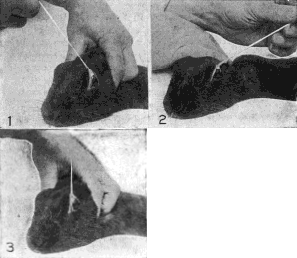
Fig. 15. Demonstration Upon the Eye of a Rabbit That the Production of Refractive Errors Is Dependent Upon the Action of the External Muscles. The String Is Fastened to the Insertion of the Superior Oblique and Rectus Muscles
No. 1.—Backward pull. Myopia is produced.
No. 2.—Forward pull. Hypermetropia is produced.
No. 3.—Upward pull in the plane of the iris. Mixed astigmatism is produced.
After one or both of the obliques had been cut across, or after they had been paralyzed by the injection of atropine deep into the orbit, accommodation could never be produced by electrical stimulation; but after the effects of the atropine had passed away, or a divided muscle had been sewed together, accommodation followed electrical stimulation just as usual. Again when one oblique muscle was absent, as was found to be the case in a dogfish, a shark and a few perch, or rudimentary, as in all cats observed, a few fish and an occasional rabbit, accommodation could not be produced by electrical stimulation. But when the rudimentary muscle was strengthened by advancement, or the absent one was replaced by a suture which supplied the necessary countertraction, accommodation could always be produced by electrical stimulation.
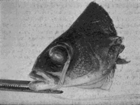
Fig. 16. Demonstration Upon the Eye of a Fish That the Production of Myopic and Hypermetropic Refraction Is Dependent Upon the Action of the Extrinsic MusclesSuture tied to the insertion of the superior rectus muscle. By means of strong traction upon the suture the eyeball is turned in its socket, and by tying the thread to a pair of fixation forceps which grasp the lower jaw, it is maintained in this position. A high degree of mixed astigmatism is produced, as demonstrated by simultaneous retinoscopy. When the superior oblique is divided the myopic part of the astigmatism disappears, and when theinferior rectus is cut the hypermetropic part disappears, and the eye becomes normal—adjusted for distant vision—although the same amount of traction is maintained. It is evident that these muscles are essential factors in the production of myopia and hypermetropia.
After one or both of the oblique muscles had been cut, and while two or more of the recti were present and active,3 electrical stimulation of the eyeball, or of the nerves of accommodation, always produced hypermetropia, while by the manipulation of one of the recti, usually the inferior or the superior, so as to strengthen its pull, the same result could be produced. The paralyzing of the recti by atropine, or the cutting of one or more of them, prevented the production of hypermetropic refraction by electrical stimulation; but after the effects of the atropine had passed away, or after a divided muscle had been sewed together, hypermetropia was produced as usual by electrical stimulation.
It should be emphasized that in order to paralyze either the recti muscles, or the obliques, it was found necessary to inject the atropine far back behind the eyeball with a hypodermic needle. This drug is supposed to paralyze the accommodation when dropped into the eyes of human beings or animals, but in all of my experiments it was found that when used in this way it had very little effect upon the power of the eye to change its focus.
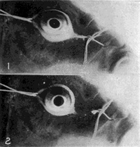
Fig. 17.
No. 1.—Production of mixed astigmatism in the eye of a carp by pulling strings attached to the conjunctiva in opposite directions. Note the oval shape of the front of the eyeball.
No. 2.—With the cutting of the strings the eyeball returns to its normal shape, and the refraction becomes normal.
Astigmatism was usually produced in combination with myopic or hypermetropic refraction. It was also produced by various manipulations of both the oblique and recti muscles. Mixed astigmatism, which is a combination of myopic with hypermetropic refraction, was always produced by traction on the insertion of the superior or inferior rectus in a direction parallel to the plane of the iris, so long as both obliques were present and active; but if either or both of the obliques had been cut, the myopic part of the astigmatism disappeared. Similarly after the superior or the inferior rectus had been cut the hypermetropic part of the astigmatism disappeared. Advancement of the two obliques, with advancement of the superior and inferior recti, always produced mixed astigmatism.
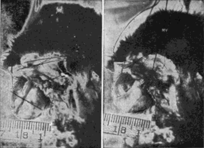
Fig. 18. Demonstration Upon the Eyeball of a Rabbit That the Obliques Lengthen the Visual Axis in Myopia
R, rest. The eyeball is of normal length and emmetropic—that is, perfectly adjusted for distant vision. My, myopia. The pull of the oblique muscles has been strengthened by advancement and the retinoscope shows that myopia has been produced. It can easily be noted that the eyeball is longer. It was impossible to avoid some movement of the head between the taking of the two pictures as a result of the manipulation of the strings, butthe rule shows that the focus of the camera was not appreciably changed by such movements.
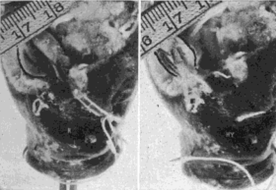
Fig. 19. Demonstration Upon the Eye of a Carp That the Recti Shorten the Visual Axis in Hypermetropia
R, rest. The eyeball is of normal length and emmetropic. Hy, hypermetropia. The pull of the external and internal recti has been strengthened by advancement, and the retinoscope shows that hypermetropia has been produced. It may easily be noted that the eyeball is shorter. The rule shows that the focus of the camera was not appreciably changed between the taking of the two pictures.
Eyes from which the lens had been removed, or in which it had been pushed out of the axis of vision, responded to electrical stimulation precisely as did the normal eye, so long as the muscles were active; but when they had been paralyzed by the injection of atropine deep into the orbit, electrical stimulation had no effect on the refraction.
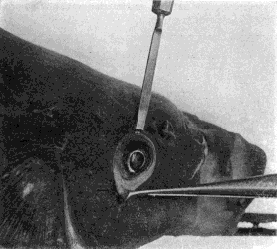
Fig. 20. Lens Pushed Out of the Axis of Vision
In this experiment on the eye of a carp the lens was pushed out of the axis of vision. Accommodation took place after this displacement just as it did before. Note the point of the knife in the pupil in front of the lens.
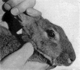
Fig. 21. Rabbit With Lens Removed
The animal was exhibited at a meeting of the Ophthalmological Section of the American Medical Association, held in Atlantic City, and was examined by a number of ophthalmologists present, all of whom testified that electrical stimulation of the eyeball produced accommodation, or myopic refraction, precisely as in the normal eye.
In one experiment the lens was removed from the right eye of a rabbit, the refraction of each eye having first been tested by retinoscopy and found to be normal. The wound was then allowed to heal. Thereafter, for a period extending from one month to two years, electrical stimulation always produced accommodation in the lensless eye precisely to the same extent as in the eye which had a lens. The same experiment with the same result was performed on a number of other rabbits, on dogs and on fish. The obvious conclusion is that the lens is not a factor in accommodation.
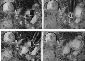
Fig. 22. Experiment Upon the Eye of a Cat Demonstrating That the Fourth Nerve, Which Supplies Only the Superior Oblique Muscle, Is Just as Much a Nerve of Accommodation As the Third, and That the Superior Oblique Muscle Which It Supplies Is a Muscle of Accommodation
No. 1.—Both nerves have been exposed near their origin in the brain, and a strip of black paper has been inserted beneath each to render it visible. The fourth nerve is the smaller one The superior oblique muscle has been advanced by a tucking operation, as this muscle is always rudimentary in cats,and unless its pull is strengthened accommodation cannot be produced in these animals. Stimulation of either or both nerves by the faradic currentproduced accommodation.
No. 2.—When the fourth nerve was covered with cotton soaked in a normal salt solution, the application of the faradic current to the cottonproduced accommodation. When the cotton was soaked in a one per cent solution of atropine sulphate in a normal salt solution, such applicationproduced no accommodation, but stimulation of the third nerve did produce it.
No. 3. When the third nerve was covered with cotton soaked in a normal salt solution, the application of the faradic current to the cotton producedaccommodation. When the cotton was soaked with atropine sulphate in a normal salt solution, such application produced no accommodation, but thestimulation of the fourth nerve did produce it.
No. 4.—When both nerves were covered with cotton soaked in atropine sulphate in a normal salt solution, the application of electricity to the cottonproduced no accommodation. When the parts had been washed with a warm salt solution electrical stimulation of either nerve always producedaccommodation. The nerves were alternately covered with the atropine-soaked cotton and then washed with the warm saline solution for an hour theelectricity being applied in each condition with invariably the same result. Accommodation could never be produced by electrical stimulation when thenerves were paralyzed with the atropine, but always resulted from the stimulation of either or both when they had been washed with the saltsolution. The experiment was performed with the same results on many rabbits and dogs.
In most text-books on physiology it is stated that accommodation is controlled by the third cranial nerve, which supplies all the muscles of the eyeball except the superior oblique and the external rectus; but the fourth cranial nerve, which supplies only the superior oblique, was found in these experiments to be just as much a nerve of accommodation as the third. When either the third or the fourth nerve was stimulated with electricity near its point of origin in the brain accommodation always resulted in the normal eye. When the origin of either nerve was covered with a small wad of cotton soaked in a two per cent solution of atropine sulphate in a normal salt solution, stimulation of that nerve produced no accommodation, while stimulation of the unparalyzed nerve did produce it. When the origin of both nerves was covered with cotton soaked in atropine, accommodation could not be produced by electrical stimulation of either or both. When the cotton was removed and the nerves washed with normal salt solution, electrical stimulation of either or both produced accommodation just as before the atropine had been applied. This experiment, which was performed repeatedly for more than an hour by alternately applying and removing the atropine, not only demonstrated clearly what had not been known before, namely, that the fourth nerve is a nerve of accommodation, but also demonstrated that the superior oblique muscle which is supplied by it is an important factor in accommodation. It was further found that when the action of the oblique muscles was prevented by dividing them, the stimulation of the third nerve produced, not accommodation, but hypermetropia.
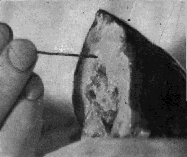
Fig. 23. Pithing a Fish Preparatory to Operating Upon Its Eyes
The object of this operation is to secure greater relaxation of the muscles of the eyes and head, which would work for hours, without external stimulus, if the brain cells were not destroyed by the probe.
In all the experiments all sources of error are believed to have been eliminated. They were all repeated many times and always with the same result. They seemed, therefore, to leave no room for doubt that neither the lens nor any muscle inside the eyeball has anything to do with accommodation, but that the process whereby the eye adjusts itself for vision at different distances is entirely controlled by the action of the muscles on the outside of the globe.
1. The refractive system.
2. On the Anomalies of Accommodation and Refraction of the Eye, p. 22.
3. In many animals, notably in rabbits, the internal and external recti are either absent or rudimentary, so that, practically, in such cases, there are only two recti, just as there are only two obliques. In others, as in manyfish, the internal rectus is negligible.
| Eyes carePhysicianBate's booksLaser corre.Blues under eyesburning in the eyesanother diseasesMedical mistery Naturally eyesight correction. No laser eye surgery. Restore eyesight. Vision correction. Los conflictos de alquiler requieren respuestas legales claras. Deja tu caso en manos de Abogados especialistas en alquileres Madrid En VMS Abogados actuamos con rapidez para proteger tus intereses. | Bigwin br | |