


Strengthening the Eyes
by Bernarr A. MacFadden, Strengthening the Eyes; A System of Scientific Eye Training.
CHAPTER II
The Anatomy of the Human Eye
A SIMPLIFIED exposition of the structure of this wonderful organ is imperative in a course of this character in order that the pupil may understand the terms which follow, but only a very brief summary can be attempted. For a more detailed account of the structure and physiology of the eye the student is referred to larger works.
Normally, the eyeball is nearly spherical in shape, and has three membranes, or coats, and three humors. The external coat is a thin, tough membrane, which maintains the form of the ball; it is called the sclerotic, and forms what is known as the "white of the eye," and includes the anterior four-fifths of the outer coat; the anterior one-fifth is the cornea, a transparent disk joined to the sclerotic somewhat as a watch-glass is set in its case. It can be plainly seen by looking at the eye sideways.
The next coat which lies against the inner surface of the sclerotic and is very vascular, is called the choroid.
The choroid is composed of a network of blood-vessels, and is lined with a layer of pigment cells, whose duty it is to absorb an excess of light.
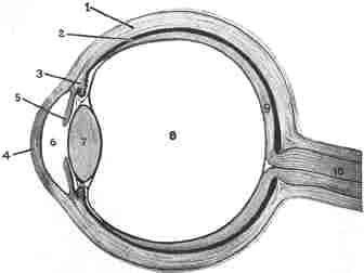
Horizontal and sectional view of the structure of the eye. (1) Sclerotic coat; (2) Choroid coat; (3) Ciliary body; (4) Cornea—the "watch glass" in front of the eyeball; (5) The iris; (6) Anterior chamber, containing aqueous humor; (7) Crystalline lens; the pupil is between 6 and 7; (8) Vitreous Humor, filling the eyeball; (9) Retina; (10) Optic nerve.
The iris, which forms a thin curtain behind the cornea, gives the eye its special color and to a large extent its beauty. The color of the iris in newly born babies is blue, and the differing colors which come later in life are due to the addition of a greater or lesser amount of dark pigment. The color is usually more or less in uniformity with the general coloring of the individual.
The pupil is merely an opening in the center of the iris, and appears black because of the darkness of the interior of the eye. Through it the rays of light, coming from any object, must pass. The pupil has the power of contracting or expanding under the influence of light; and certain drugs, such as opium and belladonna, cause it to contract or dilate unnaturally for long periods of time. The pupils of cats, tigers and other animals appear to shine in the dark; and for long it was thought that this phenomenon was due to some form of phosphorescence, but it is now known to be merely a reflection from the cornea.
At the junction of the iris and choroid is found a narrow band of delicate muscular fibers, called the ciliary muscle. This little muscle has been thought to play a very important part in the workings of the eye, notably in its "accommodation," and should be remem-bered, as it will be referred to repeatedly further on.
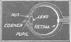
Simplified diagram, corresponding to the figure on page 8.
The retina, the third or nervous membrane, lies at the back of the eye-wall, and upon it the light-rays entering the eye are thrown or "focused." It is an exceedingly delicate and sensitive structure, liable to injury and less than one-hundredth of an inch thick. Nevertheless about ten different layers have been found within it! The outermost of these, called "Jacob's membrane," has been found to consist of minute columns arranged side by side perpendicular to the choroid, while the internal, or nerve-fiber layer, is composed of delicate nerve-fibrils forming a surface parallel to the choroid.
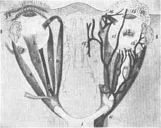
View of eyeballs from above, showing the muscles and arteries. (1) Crossing of the optic nerve; (2) Superior rectus muscle; (3) Inferior rectus muscle; (4) External rectus muscle; (5) Internal rectus muscle; (6) Superior oblique muscle (7) Inferior oblique muscle; (8) Lachrymal glands; (9) Eyelid in section; (10) Eyelid from inside; (11) Infra-orbital artery; (12) Branch to the tear gland; (13) Branch to the retina; (14) Branch to the iris; (15) Branch to the upper eyelid; (16) Branch to the eyebrow; (17) Branch to the cavity of the nose.
The optic nerve passes from the eye to the brain, and carries the nervous impulses which, in the sight-centers of the brain, are converted into the "sensation of seeing." There is evidence that the optic nerve carries impulses that result in pain, but apart from this it can carry only one kind of nerve impulse, that of sight. Hence, when it is stimulated, by whatever means, whether normally by light, or by an electric current, or by a blow, we get the impression of light or "seeing stars."
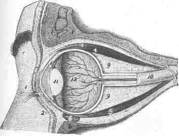
Section through the right eye. (1) Upper eyelid; (2) Lower eyelid; (3) Eyelid lifting muscle; (4) Superior rectus muscle; (5) Inferior rectus muscle; (6) Inferior oblique muscle; (7) Frontal bone; (8) Superior maxillary bone; (9) Fat; (10) Optic Nerve; (11) Crystalline lens; (12) Vitreous humor.
The three humors are the aqueous, crystalline and vitreous.
The vitreous humor occupies about four-fifths of the interior of the ball; it is colorless and transparent, and somewhat resembles a very thin jelly. It is solid enough to maintain the shape of the eye, while at the same time yielding readily under pressure. It is firm, yet elastic.
The crystalline humor, or lens, is firmer than the vitreous, but not solid, and is shaped somewhat like an ordinary magnifying glass. It grows denser with age. This also is a very important part of the eye, and will be dealt with / more fully in the discussion of accommodation ' and errors of refraction.
The aqueous humor is nearly pure water, and is contained in the space between the cornea and lens.
The orbit is the hollow cone of dense protective bone in which the eye is set. The roof of the orbit, however, is very thin, and upon this the fore-brain rests. It may be injured by a blow from beneath; and duelists are said to have selected this spot for a fatal sword-thrust. King Henry II of France was accidentally killed at a tournament by a lance point which pierced his brain through this fragile bone.
The eyebrows are formed of bone, muscle and thick skin, covered with hairs, and protect the eyes from drops of sweat, water, dirt, etc., which might otherwise find their way into them.
The eyelid is also a protective covering, composed of a layer of loose skin, and covers the eyes during sleep, when the ball is "everted," or turned upwards, also from dust, smoke, etc.
The polish and transparency of the cornea are maintained by frequent unconscious winking, which keeps its surface moist and free from dust. The mucous lining of the eyelids is always more or less moist, and is continuous with skin at the margin of the lids. After lining the inner surface of the lids it passes over to the ball, forming a loose fold, which is the only direct connection between them; hence its name, conjunctiva. It covers the front part of the sclerotic, the whole of the visible portion, and lining the walls of the tear-duct, becomes continuous with the membrane of the nose and throat, and, therefore, usually takes a part in a "cold in the head," or influenza. It is usually transparent, but may become bloodshot, or yellow, as in jaundice. Yellowness results when the coloring matter of the bile is deposited in the conjunctiva.
The opening between the lids is called the commissure, and the apparent size of the eyes depends chiefly upon the width of this space. The almond shape of Oriental eyes is due to the unusual length of the fissure between the lids, apparently increased by the absence of the plica semilunaris—the small triangle of flesh at the inner angle of the eyelids. In the Chinese, the outer angle of the commissure is much higher than the inner, giving the cleft an obliquity upwards and outwards. This has a marked effect upon the whole expression of the face.
The lachrymal apparatus consists of the gland for secreting tears and the passages for draining them off. The "tear glands" are situated just above the outer angle of the eye, and a number of small ducts carry the tears, when secreted, to the eye itself. After passage across the surface of the eye, the tears are taken up by passages, which commence near the inner angle of the eye, and are conducted into the nose. The tubes carrying the tears to the nasal passages are called the lachrymal canals. Two tiny holes or outlets permit the tears to enter these canals from the surface of the eye. Tears are usually drained off in this manner, and only "overflow" and drop off the lids when the glands are excited by excessive emotion or by local irritation. "Sniffing" is usually the first stage of a "good cry." Infants do not shed tears before the third to fourth month, and the elephant is the only one of the lower animals accused of this human weakness—statements concerning the crocodile to the contrary notwithstanding!
The eyeball is moved in various directions by a number of muscles, attached to it at the back, top, bottom and sides. It can thus be turned upwards, downwards, inwards or outwards, or may be rotated. When these muscles are uniformly relaxed or acting in unison, the eye is normal. When, from any cause, one set of muscles exerts a stronger pull than its opposites, a squint is produced. When the muscles contract excessively, they squeeze the eyeball out of shape, elongating it or the reverse. The importance of this fact we shall see when we come to the chapter devoted to errors of refraction.
| Eyes carePhysicianBate's booksLaser corre.Blues under eyesburning in the eyesanother diseasesMedical mistery Naturally eyesight correction. No laser eye surgery. Restore eyesight. Vision correction. |