


Strengthening the Eyes
by Bernarr A. MacFadden, Strengthening the Eyes; A System of Scientific Eye Training.
CHAPTER III
How We See: The Physiology and Physics of Vision
THE act of seeing is one of the greatest mysteries in the world. To the ancients it was an even greater mystery than to us, and continued to be so until the great astronomer Kepler noted the resemblance between the human eye and the camera, and demonstrated that images of external objects are formed by the organs of vision exactly as they are by the photographer's apparatus. In the eye, the rays of light, coming from the object seen, traverse the eyeball and fall upon the sensitive retina (the "plate" of the camera), from which they are conveyed to the sight-centers of the brain by the fibers of the optic nerve. That is, the impression which they have created upon the retina is so conveyed.
In order that the reader may understand the optics and physics of the eye, and of sight, it is necessary to say a few words concerning light, and reflecting and refracting bodies and surfaces.
Light is primarily given forth by self-luminous bodies, such as a candle or the sun; and everything else is reflected light. The earth reflects the rays of the sun, and this gives us our "daylight." When this reflection is cut off, dense blackness prevails. The spaces between the stars are inky black, for there is no solid body to reflect the rays coming from the sun. Rays of light are reflected when the body on which they shine sends them back. And if the light strikes a reflecting surface at an angle, the reflected angle—the so-called "angle of reflection"—is always equal to the "angle of incidence," or the direction of the shaft of light striking the object.
The white light we see is composed, as we know, of seven primary colors. Some bodies absorb some of these vibrations, and reflect others; and when this is the case such bodies are said to be colored in various ways. The color of any object is not inherent in it, but is due solely to the fact that some of the light-rays are reflected and some absorbed. Those which are reflected constitute its "color."
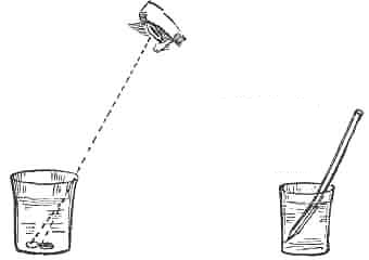
Refraction of light by the surface of water, illustrated by the coin which in an empty cup may be invisible and which appears to move upward and come within the range of vision when the cup is filled with water. Refraction is also seen in the appearance of the bending of a stick or pencil immersed in water, when looking at it from above.
When rays of light pass from one medium into another of different are bent of their previous course, which in the case of rays coming from a distance, is one of parallel lines. A simple experiment which demonstrates this is that in which a small coin is thrown into a basin of water. It appears to be in a certain place, but if an attempt be made to touch it in this place it is found to be not there at all, but somewhere else! "Appearances are deceptive." This is due to the fact that water, being of a density different from that of air, bends the rays of light and makes them take a different direction; they are, in short, "refracted." By suitable means, these light-rays can be straightened out again; they may also be refracted any number of times and in various ways.
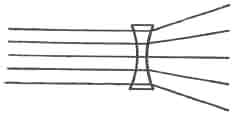
Refraction of light-rays passing through concave lens, becoming divergent.
Thus, a plain, flat sheet of glass will bend all the light-rays which pass through it at the same angle. They are neither diverged (scattered), nor brought to a point (converged). When, however, the glass is double "convex," the rays of light are brought together into a "focus." When it is double "concave," the light-rays are scattered, or diverged.
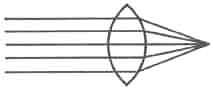
Refraction of light-rays passing through convex lens, becoming convergent.
We have seen vergent. that light-rays passing through a double convex glass or lens converge at a certain point, and if, at that point, we place a screen, we catch an image of the object from which they proceed. In the eye, precisely this phenomenon takes place. The cornea and crystalline lens focus the light-rays upon the retina at the back of the eye, which catches the image, and from this point the impression is carried along the optic nerve to the brain, as before described. When the rays are focused exactly upon this delicate surface, the impression is clear and distinct. Otherwise it is blurred, or sight may fail altogether.
All parts of the retina are not equally sensitive to visual impressions. The most sensitive portion is a little depression directly in the line of vision, called the fovea centralis, literally, the central pit. Indeed, this is the only spot which admits direct vision. In all other places it is more or less blurred. We can see only one thing at a time clearly; the rest becomes blurred and fades out as it recedes from the central point.
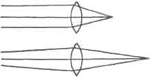
Illustrating the refraction of rays of light from distant and nearby points. The parallel rays, from a distant point, are concentrated at a point much closer to the lens than the divergent rays from a nearby point, which are focused further back. This is also demonstrated by the familiar experience of focusing light-rays in a camera.
Not far from this most sensitive spot there is a blind spot on the retina, which is unable to see anything. We have one in each eye, but the eyes are so adjusted that when one eye is blind the other sees, and vice versa. This blind spot is at the entrance of the optic nerve to the retina, and its locality can easily be seen in the diagram on the preceding page.
Illustrating the refraction of rays of light from distant and nearby points. The parallel rays, from a distant point, are concentrated at a point much closer to the lens than the divergent rays from a nearby point, which are focused further back. This is also demonstrated by the familiar experience of focusing light-rays in a camera.
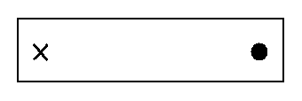
To prove the existence of the blind spot:
Close the left eye and direct the right eye to the small cross on the left hand side of the figure. Hold the page vertically before the eye, ten or twelve inches off, and then gradually bring it nearer, still keeping the gaze fixed upon the cross. The round spot will also be visible, except at a certain distance from the eye—about seven inches—when it will disappear from view. Its image falls upon the point of entrance of the optic nerve, which is incapable of perceiving light.
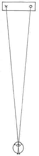
On blind spot.
In the diagram on this page is illustrated how the image is thrown upon the retina in the "blind spot" experiment. The eyeball in this diagram is proportionately small. The cross marks the spot of entrance of the optic nerve.
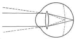
Diagram illustrating myopia or near-sightedness, rays of light from a distance (indicated by broken lines) falling in front of the retina.
We have seen that a double convex lens will focus light-rays at a certain point or distance beyond it; this distance will depend upon the degree of convexity of the lens. If it is only slightly convex, the focal point will be some distance away, while if it is very much curved, the focal point will be very near. We have only to alter the degree of convexity of the lens to insure the focusing of the rays at any desired distance (within limits).
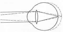
Diagram illustrating hypermetropia or far sightedness, light-rays from nearby points (indicated by broken lines) being focused behind the retina.
Now, the lens of the eye is like any other lens in this respect; and it was long ago pointed out that if it were slightly altered in shape, by means of muscular tension or otherwise, rays of light coming from different distances could be focused accurately upon the retina. It has been believed for many years that the eye adjusts itself for vision at different distances by this means, and the theory, which was accepted mainly upon the authority of Helmholtz, is the basis of the teaching in all the text books on ophthalmology today. This change of focus is called "accommodation," and is supposed to be effected by means of the ciliary muscle.
When the eye does not adjust itself properly for vision at different distances it is said to be suffering from an error of refraction. These errors, with the exception of presbyopia (old-age sight), are attributed to a wrongly-shaped eyeball (occasionally to a wrongly-shaped lens), and are believed to be either congenital (present at birth) or acquired. Presbyopia is attributed to the hardening of the lens and its consequent inability to change its shape. It is stated in all the text books on ophthalmology that these conditions are incurable and almost entirely unavoidable; but, by altering the shape of the lens, the ciliary muscle is supposed to be capable of compensating to some extent for deviations from the normal in the shape of the eyeball, and when the patient is not physically at par this is believed to impose a great strain on the eye and the nervous system.
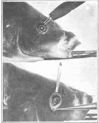
An experiment on the eye of a carp demonstrating that the lens is not a factor in accommodation. In the upper picture the eye is normal and accommodates normally when stimulated by the electrode. In the lower the lens has been pushed out of the line of sight by an instrument the point of which can be seen in the pupil. The eye is then stimulated by electricity and accommodates precisely as in the upper picture.

Production of astigmatism in the eye of a carp by changing the shape of the eyeball. In the upper picture the pull of two strings attached to the conjunctiva has made the cornea oval, thereby producing astigmatism. In the lower one the string has been cut, the cornea has resumed its natural shape and the eye is normal.
It is upon these doctrines that the treatment of refractive errors by means of glasses is based. So long as the lens and the ciliary muscle worked, and the eyeball was of the normal shape (or the lens and ciliary muscle were capable of compensating for its deficiencies), the light -rays, we have been told, were focused accurately upon the retina, and the eye required no help. But when this was no longer possible or easy, for any reason, then an extra lens was prescribed to assist in bending the light-rays so that they should be properly focused. This often improved the vision and seemed to relieve strain. In other cases it did neither, and the patient wandered hopelessly from one specialist to another in the vain search for relief.
As we shall see, however, when we come to the further discussion of accommodation and refractive errors (Chapter VI), these theories are erroneous.
| Eyes carePhysicianBate's booksLaser corre.Blues under eyesburning in the eyesanother diseasesMedical mistery Naturally eyesight correction. No laser eye surgery. Restore eyesight. Vision correction. |